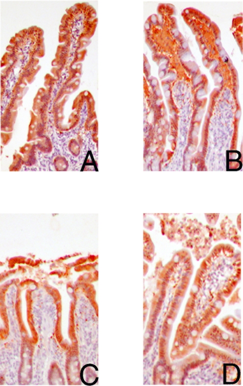
Figure 2. Immunolocalization of I-FABP in red (3-amino-9-ethylcarbazole, AEC) (100×) in the control jejunum not subjected to ischemia-reperfusion (A) shows an abundant cytosolic presence of I-FABP in the epithelial cells of the upper half of the villus.
Upon 30 minutes ischemia (B), cytosolic I-FABP staining is decreased in mature enterocytes with abundant staining in the subepithelial spaces. A decreased cytosolic staining is still observed after 25 minutes reperfusion (C). Within 60 minutes reperfusion, I-FABP cytosolic positive cells are part of the resealed epithelial barrier (D), while shedded I-FABP containing enterocytes are found in the debris in the lumen.

