
Fig. 2.
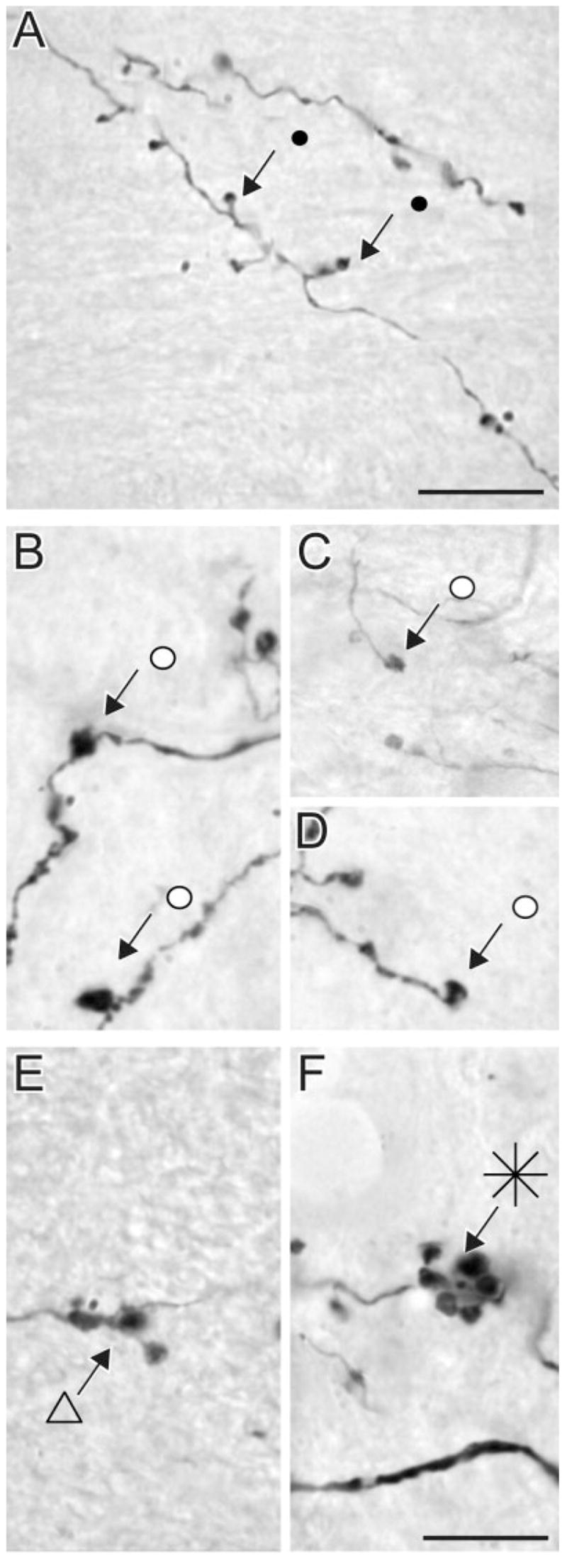
High-magnification photomicrographs of terminal types in the LPl-1 originating from area PMLS. Type I terminals area small boutons linked to small-diameter axons by short stalks (A). Singleton terminals form swellings along the axon (B) or single boutons at the end of long axon side branches (C,D). Type II terminals tend to group in clusters of endings of intermediate complexity (E) or form more complex rosette-like structures (F). Scale bars = 10 μm (applies to B–F).