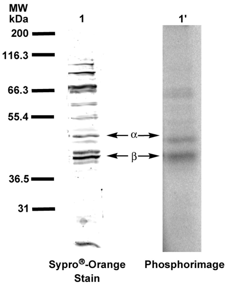
Figure 7.

Analysis of crude hPGGTase-I with [32P]3b by SDS-PAGE. Lanes 1 and 1′ contain samples of crude cell lysate after ion-exchange chromatography, irradiated at 350 nm in the presence of [32P]6. Lane 1 shows protein identified with Sypro®-orange staining. Lane 1′shows radiolabeled protein identified with phosphorimaging.
