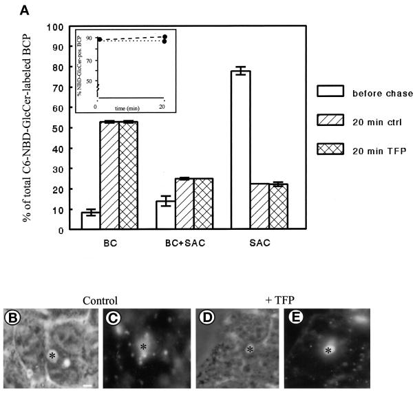
Figure 3.
Effect of TFP on transport of C6-NBD-GlcCer from SAC. C6-NBD-GlcCer was accumulated into SAC as described in MATERIALS AND METHODS (also cf. van IJzendoorn and Hoekstra, 1998). After abolishing the remaining BC-associated NBD fluorescence with sodium dithionite, cells were treated with HBSS (control) or 20 μM TFP at 4°C for 30 min. Then cells were washed and kept in HBSS at 4°C until use (<30 min; t = 0, before chase), or alternatively, cells were warmed to 37°C and incubated in back-exchange medium for 20 min. Cells were then rapidly cooled and kept on ice until use (<30 min). (A) Semiquantitative analysis of C6-NBD-GlcCer transport within the BCP. The percentage of C6-NBD-GlcCer-labeled BCP (inset; dotted line, control; dashed line, TFP treated) and the distribution of the BCP-associated NBD-GlcCer (i.e., BC, SAC, or both) were determined as described in MATERIALS AND METHODS. Data are expressed as mean ± SEM of at least three independent experiments, carried out in duplicate. (B–E) Photomicrographs illustrating the distribution of the lipid analogue in the BCP after a chase from SAC in nontreated (B and C) and TFP-treated (D and E) cells (B and D, phase-contrast to C and E, respectively; asterisk, apical, bile canalicular PM). Bar, 5 μm.

