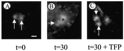
Figure 6.
Effect of TFP on SAC-to-BC transport of TxR-labeled dIgA. Cells that stably express the polymeric immunoglobulin receptor (van IJzendoorn and Hoekstra, 1998) were washed and incubated with excess asialofetuin to saturate asialoglycoprotein receptors. Subsequently, cells were incubated with 50 μg/ml TxR-dIgA at 4°C for 1 h. After rinsing the cells to remove nonbound ligand, cells were warmed to 37°C and incubated for 15 min to allow internalization and partial transcytosis. Then, cells were rapidly cooled to 18°C and further incubated for 90 min. (A) Basolateral PM-derived TxR-dIgA has accumulated in SAC (arrows). (B) After a subsequent incubation at 37°C, TxR-labeled both SAC and BC, indicative of transport to BC. However, in the presence of TFP, SAC-to-BC transport of TxR-IgA was severely inhibited. Most of the fluorescently labeled dIgA was still in SAC alone (arrows) with little if any TxR-dIgA transported to BC. A semiquantitative analysis is presented in Table 1. Asterisks, apical, bile canalicular PM. Bar, 10 μm.

