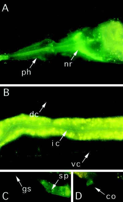
Figure 3.
Localization of a GFP-dynamin chimera under control of the dyn-1 promoter. (A) Close-up of the head region, showing that GFP-dynamin is concentrated in the nerve ring (nr). GFP-dynamin is also detected along the outside of the pharynx (ph). The yellow or orange staining of the gut is largely due to autofluorescence of gut granules. (B) Close-up of a midsection of the body, showing that the fluorescence of GFP-dynamin appears punctate along the ventral nerve cord (vc) and along the dorsal nerve cord (dc). GFP is also detectable along apical surface of the intestinal cells (ic). Some of the intestinal GFP may have been masked by the autofluorescence of gut granules (yellow-orange staining). (C) Close-up of a midsection of the body, showing fluorescence of GFP-dynamin in a spermatheca (sp) and along the gonadal sheath (gs). (D) Close-up of a midsection of the body, showing fluorescence of GFP-dynamin in a coelomocyte (co).

