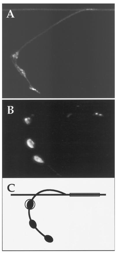
Figure 5.
Green fluorescence concentrated in presynaptic varicosities of ALM neurons. (A) Distribution of GFP near the distal end of an ALM neuron. This is a close-up of the nerve ring of a transgenic worm containing the mec-7::GFP construct. A segment of the ALM axonal process is shown at the top. The branch, which enters the nerve ring, is shown making a curve toward the bottom. The synaptic clusters are visible as three patches of fluorescence along the branch. The image is a composite in which a stack of confocal sections was merged to visualize the curved branch of the ALM process. (B) Distribution of a GFP-dynamin chimera in part of an ALM neuron. This is a close-up of the nerve ring of a transgenic worm containing the mec-7::GFP::Dynamin construct. Almost all fluorescence is concentrated in the three patches corresponding to clusters of synapses. (C) Diagram of an ALM process, with a circle around one of the clusters of synapses and a box along the axonal process depicting the areas chosen to quantitate the degree of localization. The measurements were all conducted with the same surface area (200 pixels of images taken with the same magnification).

