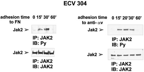
Figure 3.
Time course of adhesion-dependent JAK2 tyrosine phosphorylation. Serum-deprived ECV304 cells were detached with 10 mM EDTA and plated on dishes coated with FN (left panel) or antibodies to αv (anti-αv) (right panel) for the indicated times, kept in suspension (0 on FN) or plated on PL (0 on anti-αv). Cell extracts were prepared and subjected to immunoprecipitation (IP) with an anti-JAK2 antiserum. Proteins were electrophoretically transferred to a nitrocellulose filter that was immunoblotted (IB) with an anti-phosphotyrosine antibody (Py) (upper panels) and reprobed with an anti-JAK2 antiserum (lower panels). The position of the tyrosine-phosphorylated JAK2 is indicated. Similar results were obtained in three individual experiments.

