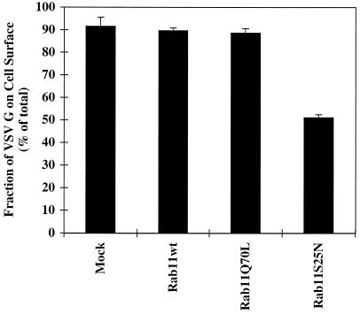
Figure 5.
Cell surface delivery of VSV G protein is reduced in cells expressing the dominant negative rab11S25N mutant. BHK cells were transfected with VSV G together with wild-type (wt) rab11, rab11Q70L, or rab11S25N. At 5 h after transfection, cells were metabolically labeled for 10 min. After a chase period of 120 min, cell surface delivery of the VSV G protein was monitored by surface biotinylation as described in MATERIALS AND METHODS. Each column represents the mean ± SEM of triplicate samples from one of three independent experiments.

