
Figure 5. SerpinB2-deficiency reduces susceptibility to Rb-dependent skin carcinogenesis.
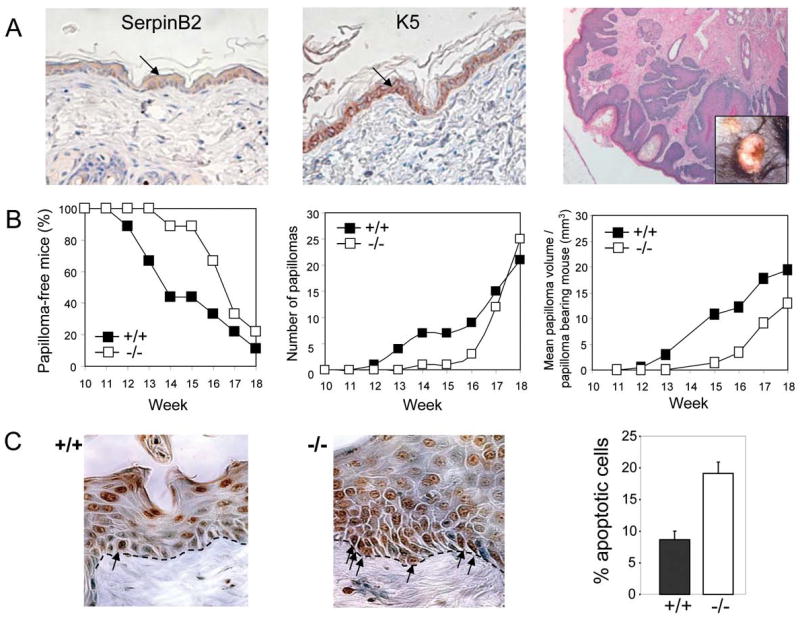
A: Histopathology of representative papillomas. In normal adult mouse epidermis, SerpinB2 is present in suprabasal and basal keratinocytes (left), where it co-localizes with basal K5 (middle), a predominant keratin of proliferating basal keratinocytes. (right) Hemotoxylin & eosin stained section of a representative SerpinB2+/+ papilloma. Inset shows gross appearance.
B: SerpinB2 deficiency reduces susceptibility to tumor formation during skin carcinogenesis. SerpinB2+/+ (n=9) and SerpinB2-/- mice (n=9) were treated with DMBA/PMA as per experiment 2 and the onset of papilloma formation followed with time. There was a significant delay in the onset of papillima incidence in the SerpinB2-/- animals (left panel). Tumor multiplicity (middle panel) represents the total number of papillomas per genotype (n=9). The mean papilloma volume per papilloma bearing mouse is represented in the right panel. Once formed, papillomas did not produce SerpinB2, and SerpinB2 deficiency had no effect on the rate of papilloma progression (data not shown).
C: Increased apoptosis in SerpinB2 deficient hyperplastic skin lesions. TUNEL staining of apoptotic keratinocytes in the basal layer of SerpinB2+/+ (left panel) and SerpinB2-/- (middle panel) epidermis. Arrows indicate TUNEL positive basal keratinocytes localized along the basement membrane (dotted line). Percentage of apoptotic cells in the basal layer (right panel). The number of TUNEL positive cells relative to the total number of cells along the basement membrane were counted. Data represents specimens from 4 mice of each genotype (2 sections per mouse) (bars, ± SD; P ≤0.001, Students t test).