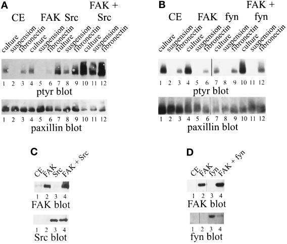
Figure 3.
Cell adhesion-dependent tyrosine phosphorylation of paxillin. (A) CE cells (lanes 1–3) or CE cells expressing RCAS A FAK (lanes 4–6), RCAS B Src (lanes 7–9), or RCAS B Src and RCAS A FAK (lanes 10–12) were analyzed. Paxillin was immunoprecipitated from lysates of cells in culture (lanes 1, 4, 7, and 10), cells held in suspension (lanes 2, 5, 8, and 11), or cells plated onto fibronectin (lanes 3, 6, 9, and 12). The immune complexes were blotted for phosphotyrosine (top panel) or paxillin (bottom panel). Expression of FAK and Src was verified by Western blotting (C). (B) CE cells (lanes 1–3) or CE cells expressing RCAS A FAK (lanes 4–6), RCAS B fyn (lanes 7–9), or RCAS B fyn and RCAS A FAK (lanes 10–12) were analyzed. Paxillin was immunoprecipitated from lysates of cells in culture (lanes 1, 4, 7, and 10), cells held in suspension (lanes 2, 5, 8, and 11), or cells plated onto fibronectin (lanes 3, 6, 9, and 12). The immune complexes were blotted for phosphotyrosine (top panel) or paxillin (bottom panel). Expression of FAK and fyn was verified by Western blotting (D).

