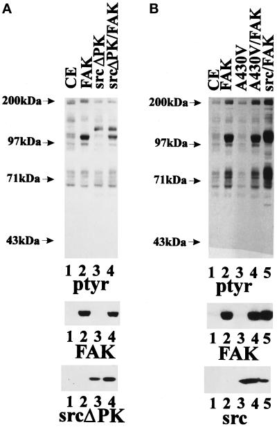
Figure 4.
Catalytically inactive pp60c-src fails to synergize with FAK. (A) Twenty-five micrograms of whole-cell lysate from CE cells (lane 1) or CE cells expressing RCAS B FAK (lane 2), RCAS A SrcΔPK (lane 3), or RCAS B FAK + RCAS A SrcΔPK (lane 4) were analyzed by Western blotting using phosphotyrosine antibodies (top panel), mAb 2A7 to detect FAK (middle panel), or mAb EC10 to detect SrcΔPK (bottom panel). (B) Twenty-five micrograms of whole-cell lysate from CE cells (lane 1) or CE cells expressing RCAS A FAK (lane 2), RCAS B SrcA430V (lane 3), RCAS A FAK + RCAS B SrcA430V (lane 4), or RCAS A FAK + RCAS B c-src (lane 5) were analyzed by Western blotting using phosphotyrosine antibodies (top panel), mAb 2A7 to detect FAK (middle panel), or mAb EC10 to detect pp60c-src (bottom panel).

