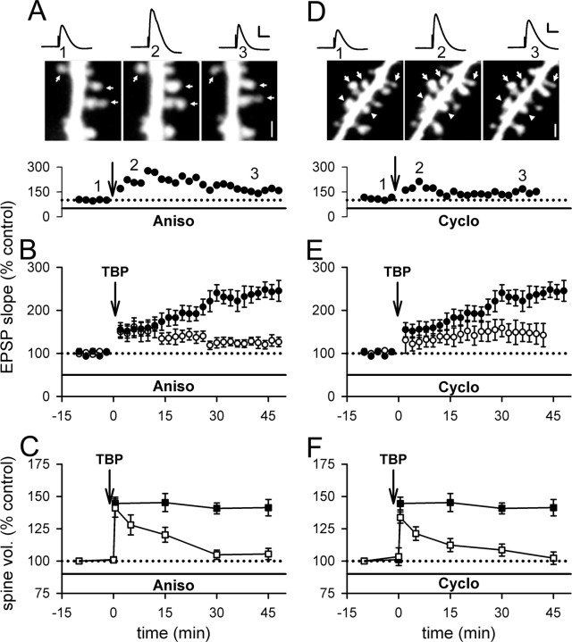
Figure 6.
Inhibiting protein synthesis reduces LTP and destabilizes spine expansion triggered by TBP. A, A representative experiment showing that EPSPs increased after TBP but decayed back toward baseline; spine expansion occurred but collapsed with time, in a neuron incubated with aniso. The sample EPSP traces and spine images were acquired at the time indicated on the graph below the images. Scale bar, 1 μm. Calibration: 5 mV, 50 ms. B, In neurons treated with aniso, TBP triggered an initial enhancement but no gradual increase in EPSPs with time (open circles). LTP in control neurons was shown in filled circles. C, The initial expansion of spines was unaffected by aniso treatment but the expansion was not sustained (open squares). Spine expansion in control neurons was shown in filled squares. D, A sample experiment showing that internal loading of cyclo led to similar changes in LTP and spine changes as in aniso-treated neurons. Scale bar, 1 μm. Calibration: 5 mV, 50 ms. E, Compared with control LTP (filled circles), the slow rising component of LTP was absent although the initial enhancement was intact (open circles) in neurons loaded with cyclo. F, Internal loading of cyclo did not affect the occurrence of spine expansion but inhibited its persistence (open squares). Spine expansion in control neurons is shown in filled squares. Error bars indicate SEM.

