
Fig. 3. Antagonism by PG13 peptide.
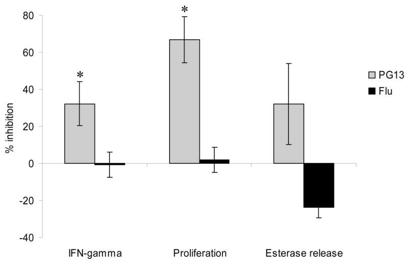
In all of the assays the AC-25 clone was stimulated with autologous B-LCL pulsed for one hour with a suboptimal concentration of agonist peptide PP16, 0.1 μg/ml for IFN-γ secretion and proliferation and 1.6 μg/ml for serine esterase release. Cells were then washed 10 μg/ml of antagonist peptide PG13 or control influenza matrix peptide PLKAEIAQRLEDV (Flu) was added. The control peptide binds DR1 but does not activate the HIV-specific clone. The left panel shows IFN-γ secretion in an intracellular cytokine staining assay with at least 50,000 events collected/condition. The middle panel shows proliferation by CFSE dilution. The right panel shows serine esterase release in the supernatant 4 hours after stimulation. All experiments were performed at least twice. * p < 0.05, PG13 vs. Flu peptide epitope.