
Figure 5. Assessment of epileptogenesis 3 weeks after intraamygdaloid KA.
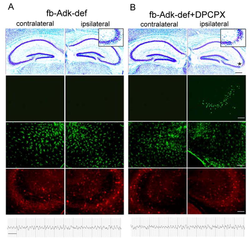
Representative micrographs and EEGs taken from the hippocampal formation of fb-Adk-def mice (A), and fb-Adk-def mice pretreated with 1mg/kg i.p. DPCPX (B), 3 weeks after intraamygdaloid injection KA. 1st row: Cresyl violet stained section showing the characteristic ipsilateral SE induced CA3 lesion in fb-Adk-def/DPCPX mice (B, asterisk), but not in fb-Adk-def mice (A). Insets show enlarged ipsilateral CA3. 2nd row: TUNEL staining showing ipsilateral CA3-selective cell loss only in fb-Adk-def/DPCPX mice. 3rd row: GFAP immunofluorescence staining showing lack of astrogliosis in fb-Adk-def and fb-Adk-def/DPCPX mice. 4th row: ADK immunofluorescence staining of the same specimen. 5th row: Representative intrahippocampal EEG recordings taken from CA3 recordings showing protection from the development of spontaneous seizures in fb-Adk-def and fb-Adk-def/DPCPX mice. Scales: black bar: 300 μm; white bar: 75 μm; EEG: 10 sec.