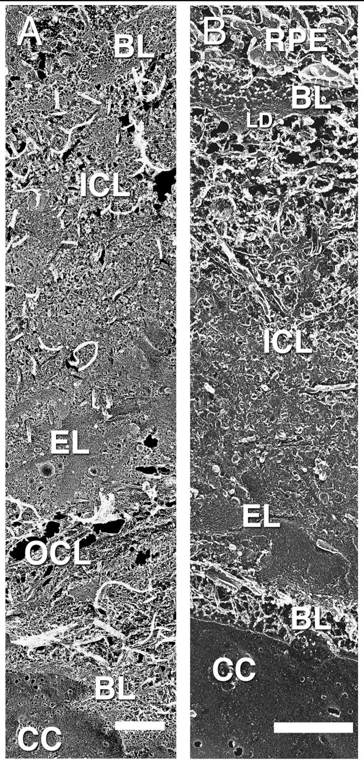
FIGURE 1.
Replica showing macular (A) and peripheral (B) BrM from the RPE at the top of the figures to the choriocapillaris on the bottom. Basal lamina (BL), inner collagenous layer (ICL), elastic layer (EL), outer collagenous layer (OCL), choriocapillaris (CC), lamina densa (LD); 63-year-old eye. Note that panels (A) and (B) are of different magnifications, because the fracture angle is different in (A) than (B). They are included in this illustration because these replicas showed the most complete demonstration of all layers in both regions. Bars are 1 μm.

