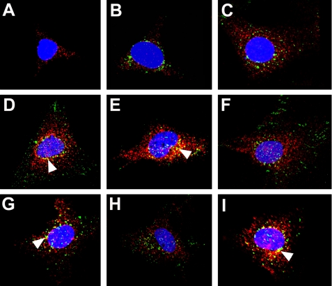
FIGURE 1.
Myotrophin co-translocates to the nucleus with p65. Neonatal rat ventricular myocytes were grown on coverslips, serum-starved, and treated with Alexa 488-labeled wild type myotrophin (green). The cells were fixed, blocked, and stained for NF-κB-p65 (red) by Alexa 568-conjugated goat anti-rabbit IgG and examined by confocal microscopy. The nuclei were stained with DAPI (blue). Co-localization of myotrophin and p65 (yellow; marked by arrowheads) is evident in certain cases. At zero time (A), p65 was observed only in the cytoplasm. Internalized Alexa 488-labeled wild type myotrophin was observed in the cytoplasm along with p65 after 5 min (B) and 10 min (C) of myotrophin treatment. Nuclear co-translocation of both Alexa labeled wild type myotrophin and p65 was observed after 20 min (D), 30 min (E), 60 min (F), 3 h (G), 4 h (H), and 16 h (I) of myotrophin treatment.

