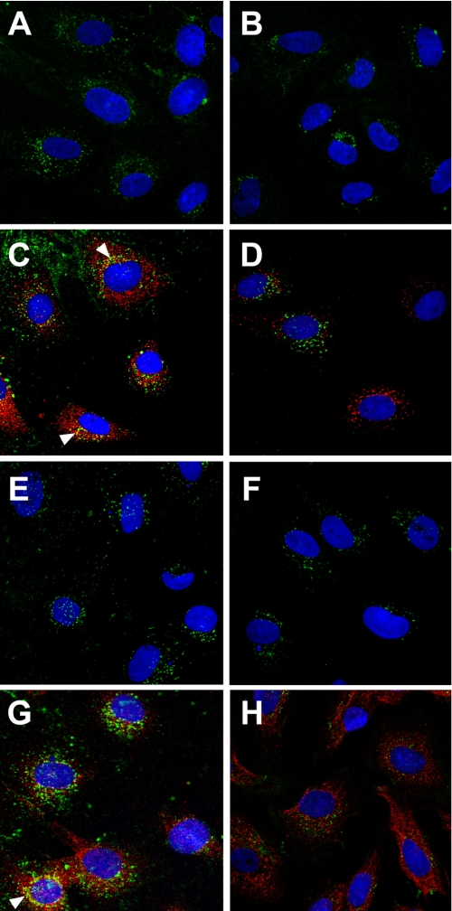
FIGURE 7.
Cellular localization of E33A mutant myotrophin protein. Neonatal rat ventricular myocytes were treated with Alexa 488 (green) labeled wild type myotrophin (left panel) or Alexa 488 (green) labeled E33A-mutant myotrophin (right panel). NF-κB-p65 co-localized (arrowheads) with Alexa 568-conjugated antibody (red) as described in the legend to Fig. 1 and in the text. After 5 min of stimulation, both the wild type (A) and E33A (B) myotrophin were observed in the cytoplasm along with p65 (C and D). After 1 h of treatment, wild type myotrophin co-translocated into the nucleus with p65 (E and G), whereas both E33A and p65 failed to co-translocate (F and H). The nuclei were stained with DAPI (blue).

