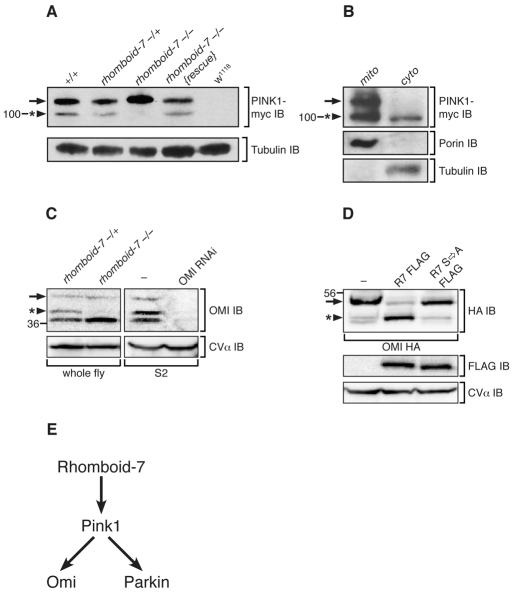
Fig. 4. Rhomboid-7 is required for cleavage of both Pink1 and Omi.
(A) Western blot analysis of Pink1-myc expression from whole fly lysates with the indicated genotypes. Pink1 appears as two forms, a long form (arrow) and a short form (arrowhead with asterisk) that is absent in rhomboid-7 mutants. Tubulin expression is used as a loading control. (B) Differential centrifugation of adult fly cells into mitochondrial (mito) and cytoplasmic (cyto) fractions. Porin and tubulin indicate the separation of mitochondria and cytosol, respectively. (C) Left panel: western blot analysis of endogenous Omi from whole fly lysates, showing a long form of Omi (arrow) and a shorter form (arrowhead with asterisk), which is absent in rhomboid-7 mutant flies. Right panel: western blot analysis of S2 cells treated with double-stranded RNA specific for Omi. The mitochondrial inner-membrane protein CV-α is used as a loading control. (D) Western blot analysis of Omi-HA expression in S2 cells with FLAG-tagged Rhomboid-7 and the Rhomboid-7 S256A catalytic mutant. (E) Schematic of the Pink1 pathway in Drosophila.

