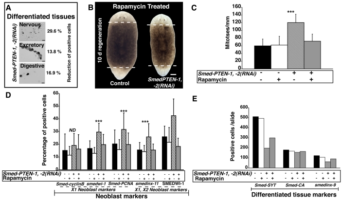
Fig. 5. PTEN-RNAi disrupts tissue maintenance and rapamycin treatment compensates for Smed-PTEN loss.
(A) Quantification of cells expressing markers of differentiated tissues. From a pool of dissociated animals, ISH was performed using probes specific to the nervous (synaptotagmin, Smed-Syt), excretory (carbonic anhydrase, Smed-CA) and digestive (smedinx-9) systems. Representative pictures of cells expressing each marker are shown (black precipitation). Cells expressing each marker were counted and the average of two independent experiments was recorded as the total number of positive cells per slide: Smed-Syt, n=608 and n=428; Smed-CA, n=109 and n=94; smedinx-9, n=142 and n=118 for control and Smed-PTEN-1, -2(RNAi) worms, respectively. Respective percentage reductions in the number of expressing cells after RNAi are shown. (B) Rapamycin treatment prevents abnormal outgrowths, lethality and rescues regenerative events in RNAi-treated animals. Worms subjected to Smed-PTEN-1, -2(RNAi) were unable to regenerate, external outgrowths were evident, and the worms failed to survive longer than 7 days post-amputation (n=27/27), whereas rapamycin-treated Smed-PTEN-1, -2(RNAi) or control worms developed anterior and posterior blastemas, and survived for more than one month after the first dsRNA injection (n=29/29). Representative images of rapamycin-treated control and Smed-PTEN-1, -2(RNAi) worms are shown. (C) Quantification of mitotic activity. Note that control and rapamycin-treated worms show similar levels of mitotic activity and that in Smed-PTEN-1, -2(RNAi) worms, proliferation was higher (***P< 0.01). However, simultaneous Smed-PTEN RNAi and rapamycin treatment prevent abnormal proliferation while keeping proliferative activity similar to control animals. Displayed values represent average ±s.d. of n≥8 worms per condition, for at least two independent replications. (D) Expression of neoblast markers in dissociated worms. An increase in the number of cells expressing each neoblast marker was always observed in Smed-PTEN-1, -2(RNAi) worms after the RNAi treatment, but was prevented with rapamycin treatment (Student’s t-test, ***P<0.001). (E) Expression of differentiated tissue markers in dissociated worms. The same pools of animals discussed in Fig. 5D were used to perform ISH with genes expressed in differentiated tissues (Smed-SYT, Smed-CA and smedinx-9). For panels (D) and (E), dissociations were performed from a pool of ten animals per group and cell numbers represent the average ± s.d. of 20 different fields per slide with an objective of 20× magnification. Total numbers of cells expressing each gene per slide are shown. ND: no difference.

