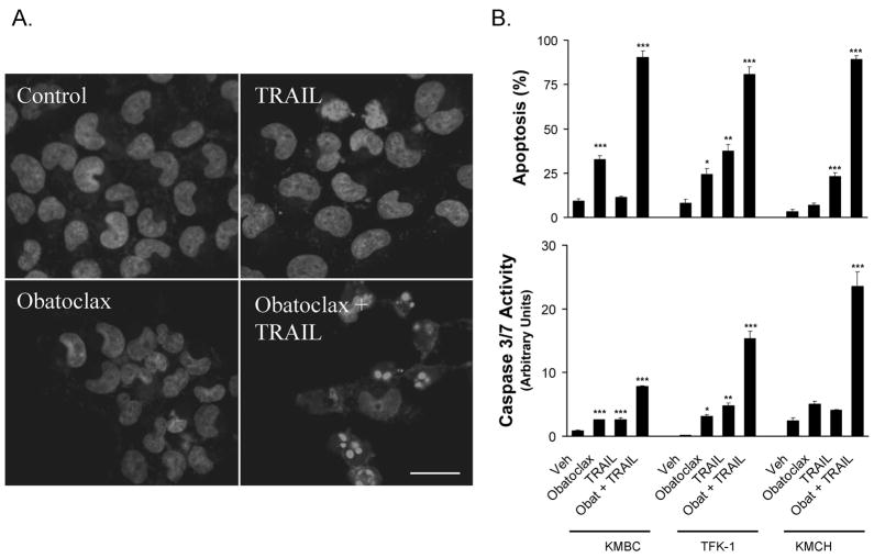
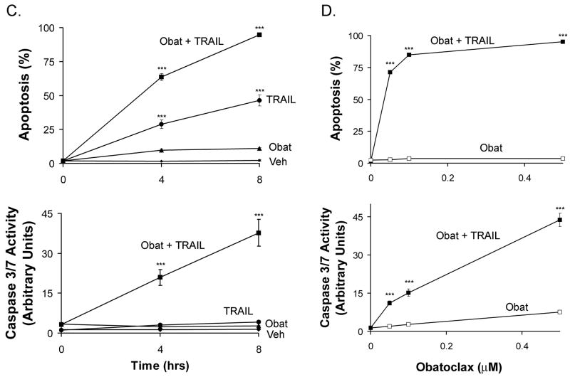
Figure 1. Obatoclax sensitizes cholangiocarcinoma cell lines to Apo2L/TRAIL killing.
Panel A: KMCH cancer cells were pretreated with 0.5 μM obatoclax or vehicle overnight, followed by obatoclax plus Apo2L/TRAIL (1 ng/mL) for the final 8 hours where indicated. Cells were fixed and stained with the nuclear dye DAPI followed by imaging by confocal microscopy. Bar = 25 micrometers. Panel B: The cholangiocarcinoma cancer cell lines KMBC, KMCH and TFK-1 were treated as in panel A, and then stained with DAPI followed by fluorescence microscopy. Apoptotic and normal nuclei were quantitated and apoptotic cells were expressed as a percent of total. Cells treated in parallel were assayed for caspase 3/7-like activity (DEVDase activity), which is expressed in arbitrary units. For statistical analysis, treatments were compared to vehicle. Panel C. Following pretreatment with diluent or obatoclax, KMCH cells were treated with or without Apo2L/TRAIL (1 ng/mL) for the indicated times followed by quantitation of apoptosis and caspase 3/7 activity. For statistical analysis, treatments were compared to vehicle at the corresponding time point.Panel D. KMCH cells were pretreated with the indicated concentration of obatoclax followed by diluent (open symbol) or Apo2L/TRAIL (1 ng/mL; closed symbol) for 8 hours. Apoptosis and caspase 3/7 activity were measured as above. All data points represent the mean ± SE of triplicate experiments. For statistical analysis, Apo2L/TRAIL plus obatoclax was compared to obatoclax alone at the corresponding concentration. For all panels, statistical significance is indicated by * = p < 0.05, ** = p < 0.01, and *** = p < 0.001.


