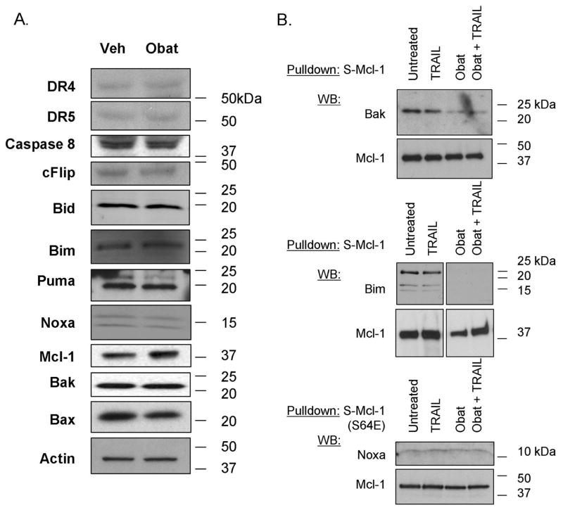
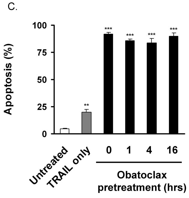
Figure 2. Obatoclax reduces Mcl-1 interaction with Bak and Bim.
Panel A: KMCH cells treated with 500 nM obatoclax for 16 hours were lysed and total protein separated by SDS-PAGE followed by immunoblot for the indicated proteins. Actin is shown as a loading control. Panel B. KMCH cells stably expressing S peptide-tagged Mcl-1 were treated overnight with medium or obatoclax followed by the addition of Apo2L/TRAIL (1 ng/mL, 8 hours) where indicated. S-peptide tagged Mcl-1 was enriched from whole cell lysates by S-protein agarose pulldown, and co-precipitating proteins examined by Western blot. The pulldown probed for Noxa (lower) used cells expressing S64E Mcl-1 (see text). Conditions are: untreated, Apo2L/TRAIL-treated, obatoclax-treated, and combined obatoclax plus Apo2L/TRAIL-treated. Images shown are cropped for clarity and the pulldown probed for Bim has been cropped to remove a redundant lane between the Apo2L/TRAIL-treated and obatoclax-treated lanes but were from the same experiment and blot. Full-length blots/gels are presented in Supplemental Figure 1. Panel C. KMCH cells were pretreated for the indicated times with medium or 0.5 M obatoclax and Apo2L/TRAIL added 8 hours before apoptosis was determined using fluorescence microscopy after DAPI staining. Statistical significance (compared to untreated) is indicated by ** = p < 0.01, and *** = p < 0.001. There was no statistical difference between cell death induced by Apo2L/TRAIL plus obatoclax pretreatment (1, 4, or 16 hours) versus Apo2L/TRAIL plus obatoclax cotreatment (0 hours pretreatment).


