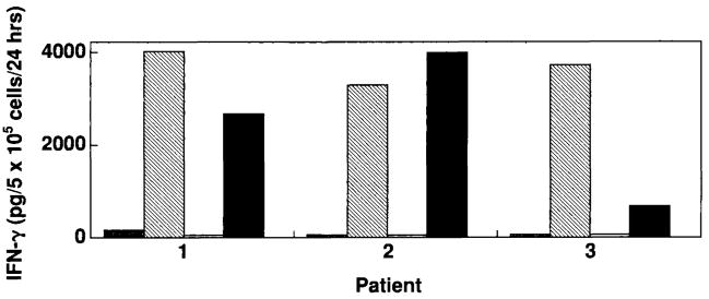
FIG. 3.
Recognition by the anti-MART-127-35 A42 cytotoxic T lymphocyte clone of endogenously processed MART-1 protein by rV-MART-1-infected dendritic cells (DC). Recognition is compared with that of MART-127-35 (1 μg/ml) pulsed DC. 105 rV-MART-1 DC (black bars) or peptide-pulsed DC (hatched bars) from three melanoma patients were coincubated with 105 A42 cells for 24 h at 37°C in 200 ml of culture medium. The efficacy of endogenous presentation by these DC was assessed by the induction of interferon-γ (IFN-γ) (pg/ml) release by A42. Negative stimulator cell controls included DC infected with an irrelevant rV encoding the sequence for the gp100 melanoma-associated antigens (white) or pulsed with the G9-209-2M peptide (gray bars). Similar results were obtained with rF-MART-1-infected DC.

