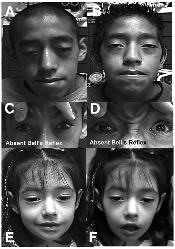
Figure 2.
External photos of three affected subjects. Cases 1 (A) and 2 (B) demonstrating ptosis, facial diplegia, and complete ophthlmoplegia. All subjects lacked Bell’s phenomenon, as seen for Cases 1 (C) and 2 (D) during attempted eyelid closure. Lower facial weekness in Case 3 is demonstrated during attempted smile (E) and facial relaxation (F).

