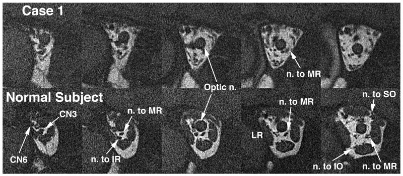
Figure 4.
Quasi-coronal MRI of deep posterior right orbit of Case 1 (top row) and a normal control subject (bottom row), illustrating hypoplasia of the deep portions of the extraocular muscles and hypoplasia or absence of the motor nerve branches to them in the affected subject. Images are in contiguous 2 mm thick planes. CN3: inferior division of the oculomotor nerve; CN6: abducens nerve; IO: inferior oblique muscle; IR: inferior rectus muscle; LR: lateral rectus muscle; MR: medial rectus muscle; SO: superior oblique muscle.

