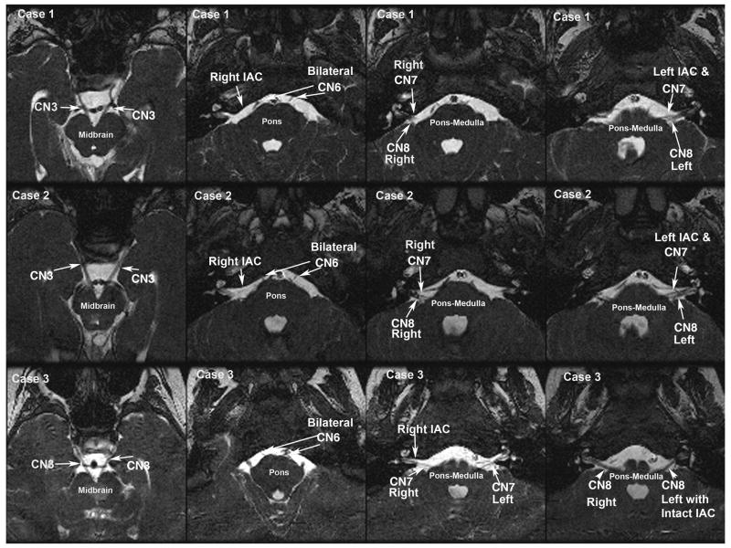
Figure 6.
Oblique axial T2 weighted MRI of brainstem from three related subjects with Möbius syndrome and ophthalmoplegia. Case 1 (top row), Case 2 (middle row), and Case 3 (bottom row) MRI panels show noncontiguous representative sections of the midbrain (left column), pons (second column), and pontine medullary junction (third and fourth columns). Cranial nerves 3, 6, 7, 8 (CN 3, CN6, CN7, CN8), and internal auditory canal (IAC) are normal and are identifed bilaterally in all subjects.

