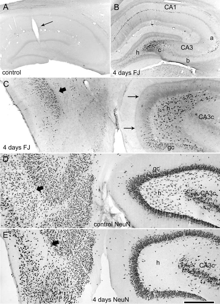
Figure 6.
Acute neurodegeneration in hippocampus and entorhinal cortex 4 days after 3 hr of perforant pathway stimulation-induced SE. (A) Coronal section of a sham control rat (implanted, no SE) showing that the presence of the hippocampal recording electrode and the control treatment (electrode implantation, stimulation for 3 hr at 0.1 Hz, halothane anesthesia, and subanesthetic urethane treatment) produced no detectable hippocampal Fluoro-Jade B (FJB) staining (grayscale, inverted image as shown in preceding figure). (B) Coronal section of a stimulated rat (same rat as in the preceding figure) showing acute injury in the dorsal hippocampus 4 days after 3 hr of perforant pathway stimulation-induced SE. Note FJB-positive cells in the hilus, area CA3a and CA3c, and area CA1. (C) Horizontal brain section from the same rat stained with FJB, showing selective degeneration of neurons in the entorhinal cortex (wide arrow), the dentate hilus (h), and the dentate granule cell layer (gc). Note also the degenerating terminals in the inner dentate molecular layer (thin arrows) that originate from the degenerating hilar mossy cells. (D) Horizontal section from a sham control animal after immunostaining for the neuronal marker NeuN. (E) A horizontal section adjacent to the section shown in (C) after NeuN immunostaining showing the loss of NeuN immunoreactivity of neurons in the hilus (h) and entorhinal cortex. Calibration bar: 1mm in A and B; 400 μm in C-E.

