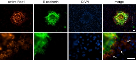
Fig. 5.
Rac1 activation in hESCs during embryo implantation. (Upper) In situ staining for active Rac1 in embryo–hESC cocultures demonstrates that cells adjacent to the implanting embryo (which is stained positive for E-cadherin) have higher levels of active Rac1 than those further away. Nuclei are counterstained with DAPI. (Lower) Higher resolution, arrows indicate cells with high levels of active Rac1. (Scale bars, 30 μm.)

