
Figure 2.
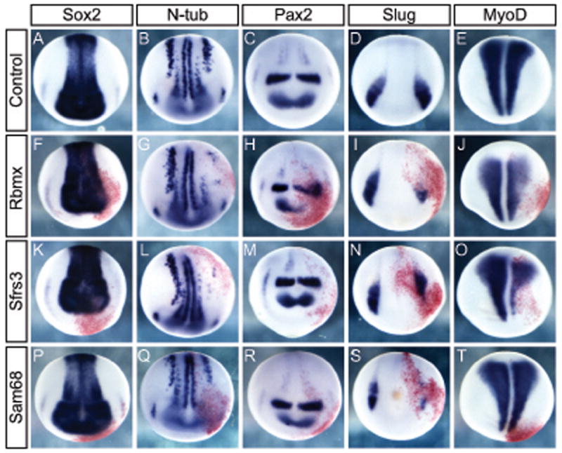
Effect of overexpression of RNA binding proteins on midneurula development. Whole mount in situ hybridization for gene expression during neurula stages on uninjected control embryos or embryos injected with 50 pg mRNA. Uninjected control embryos showing expression of Sox2 marking all neural tissue (A); N-tubulin in differentiating neurons (B); Pax2 in the forebrain, MHB, and differentiating neurons of the spinal cord (C); Slug marking neural crest (D); and MyoD marking axial mesoderm (E). RBMX mRNA injected embryos have normal neural plate morphology (F), but inhibited neural differentiation (G), and perturbed expression of Pax2 (H) and Slug (I), whereas axial mesoderm is largely unaffected (J). SFRS3 mRNA injection does not affect Sox2 expression (K), but neurogenesis is inhibited (L) though Pax2 expression in the brain (M) and Slug expression in the neural crest is intact (N). MyoD expression is reduced as response to SFRS3 (O). Sam68 injection does not alter Sox2 (P), Pax2 (R), or MyoD expression (T), but inhibit neurogenesis (Q) and Slug expression in neural crest (S). Embryos were injected with 50 pg mRNA in one cell at the 2-cell stage. Coinjected beta-galactosidase mRNA developed with red-gal was used as a lineage tracer. Embryo orientation is dorsal up, anterior facing.