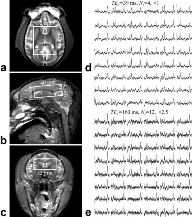
FIG. 1.

Left: (a) Axial, (b) sagittal, and (c) coronal T2-weighted MRI and typical position of the single-oblique 4.0 × 3.2 × 1.6 cm3 (AP × LR × IS) VOI in the ∼80-cm3 macaque brain. Right: Axial matrices of the real part of the spectra from the VOI over slice (a), from (d) the short TE1 = 39 ms, N1 = 4 data set and (e) the long TE2 = 165 ms, N2 = 12 data set acquisitions. All spectra are on common 3.5− 1.8 ppm and intensity scale at each TE. Long TE spectra are on a vertical scale × 2.5 times smaller than the short TE. Note the spectral resolution and SNR obtained from these isotropic (0.4 cm3) = 64-μl voxels and the substantial T2-weighting incurred from (d) to (e).
