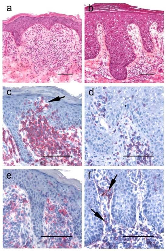
Figure 1. Immunohistochemistry results for CD3 and CD158k/e (AZ158 moAb) in representative SS and EID skin samples.

Histology and CD3 and AZ158 moAb stainings in cutaneous biopsies from the representative SS patient 8 (panels a, c, e), and from a patient with erythrodermic psoriasis (panels b, d, f) Hematoxylin and eosin-stained sections show in both samples a sub-epidermal band-like lymphocytic infiltrate associated with scattered epidermotropic lymphocytes. CD3+ T-cells (c–d), but a higher T-cell infiltrate is present in the SS sample (c), associated with Pautrier microabcesses (arrow). Lymphocytes stained with AZ158 moAb are seen in both the dermis and the epidermis in the SS sample (e), but also in a significant proportion of dermal lymphocytes (arrows) in the erythrodermic psoriasis cutaneous sample (f). Slides are shown at ×200 original magnification in panels a and b and ×400 for panels c–f. Bar = 0.1mm.
