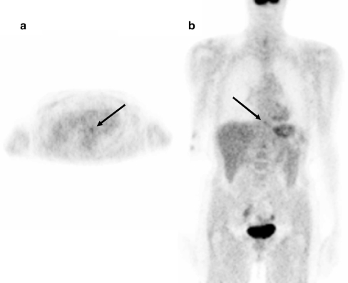
Fig. 6.
Transaxial (a) and coronal (b) [18F]FDG PET images of patient 11, displaying a rather low metabolic activity in a distal oesophageal cancer (mean SUV 2.3). Intraoperative probe readings verified a high tumour-to-background ratio of 10.4 at the surface of the oesophageal cancer. The presence of viable tumour tissue at this location was verified at histology

