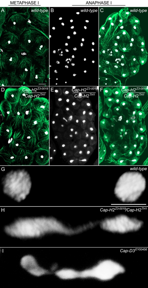
Figure 3. Chromosomes remain associated into anaphase I of Cap-H2 mutants.
Metaphase I and anaphase I morphologies were compared between wild-type and Cap-H2Z3-0019/Cap-H2TH1 mutant males. Testes were stained with DAPI and an anti-tubulin antibody to visualize DNA (white) and microtubules (green), respectively (scale bar in 3A indicates 10 µm and 5 µm in 3G). (A) Metaphase I in the wild-type. Each bivalent has congressed to the metaphase plate and appears as a cluster of DAPI stained material. (B) Anaphase I in the wild-type (DAPI only). Homologous chromosomes have segregated to daughter cells. (C) Anaphase I in the wild-type (DAPI and Tubulin merge). (D) Metaphase I from a Cap-H2Z3-0019/Cap-H2TH1 mutant male appears wild-type. (E) Anaphase I from a Cap-H2Z3-0019/Cap-H2TH1 mutant male (DAPI only). Chromatin bridges can be seen in three different segregation events. (F) Anaphase I from a Cap-H2Z3-0019/Cap-H2TH1 mutant male (DAPI and Tubulin merge). (G) Higher resolution wild-type anaphase I image showing complete segregation of homologs. (H) Anaphase I from a Cap-H2Z3-0019/Cap-H2TH1 mutant demonstrating extensive chromatin bridging due to persistent associations between chromosomes migrating to opposing poles. (I) Anaphase I bridge found from a Cap-D3EY00456 mutant.

