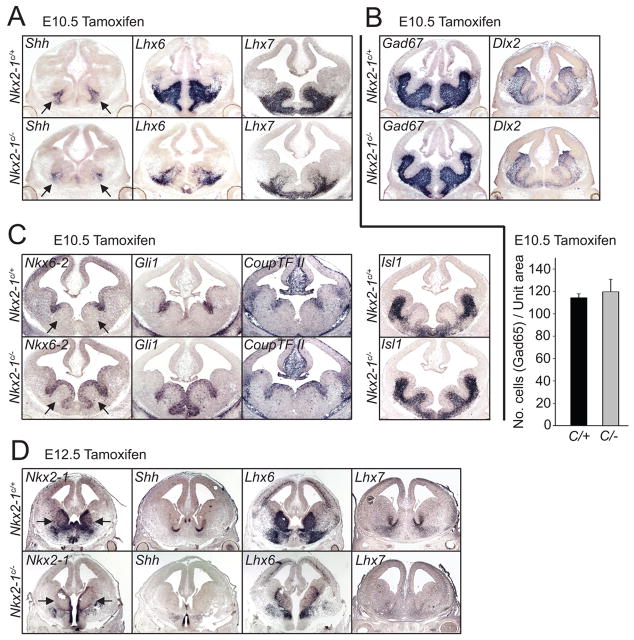
Figure 2. Conditional loss of function of Nkx2-1 at early (E10.5) and late (E12.5) time points is accompanied by changes in expression of genes involved in interneuron development.
The affects on gene expression of inducible loss of Nkx2-1 function at E10.5 (A–C) and E12.5 (D) were examined two days later using in situ hybridization of coronal sections of E12.5 (A–C) and E14.5 (D) embryos. (A) Shh, Lhx6 and Lhx7, whose expression is dependent on normal levels of Nkx2-1 in the MGE, are downregulated in Nkx2-1E10.5LOF mice. These are indicative of a reduction in the specification of the interneuron and cholinergic neuron lineages. (B) By contrast, Gad67 and Dlx2 are expressed at apparently normal levels within the subpallium, revealing that ventral GABAergic neuron development does not appear to be affected by the conditional loss of Nkx2-1 in Nkx2-1E10.5LOF mutant mice. The histogram below this photomicrograph demonstrates that the density of GAD65-positive neurons is not significantly decreased in the mutant population compared to the wild type controls. (C) The expression of Nkx6-2, Gli1 and Coup-TFII, which normally are normally expressed in both the ventricular zone of the sulcus separating the MGE and LGE, as well as portions of the CGE, are expanded throughout most the MGE ventricular zone in Nkx2-1E10.5LOF mice. Similarly, Islet1, which is normally confined to the SVZ and postmitotic regions of the LGE, expands such that it is also expressed through the SVZ of the MGE of in Nkx2-1E10.5LOF mutants. (D) Treatment of conditional Nkx2-1 mice with tamoxifen at E12.5 effectively removes Nkx2-1 expression by E14.5. Expression of Shh, Lhx6 and Lhx7 is also reduced in Nkx2-1E12.5LOF mutant mice suggesting that the loss of this gene at this age affects both the character of GABAergic and cholinergic interneuron lineages. Arrows in A, C and D indicate the position of the MGE. Error bars in the histogram of B represent SEM.

