Fig. 5.
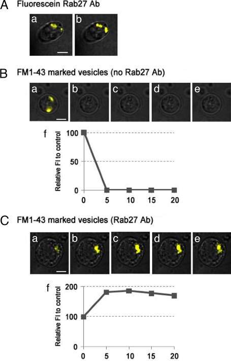
Anti-sqRab27 antibody inhibits FM1-43 dye release from synaptosomes in vitro. Fluorescein-labeled anti-sqRab27 antibody (Rab27 Ab) is loaded during synaptosome preparation and localize on synaptic vesicle profiles (Aa). After 30-min superfusion with high KCl, fluorescence vesicular profiles remain stationary in the synaptosomal images (Ab). In the case of the FM1-43 dye-loaded synaptosome without antibody, fluorescence inside the synaptosome (Ba) rapidly disappeared after high-KCl superfusion (B b–e) (n = 7). By contrast, in the unlabeled anti-sqRab27 antibody-preloaded synaptosome, FM1-43 dye remained and accumulated after high-KCl superfusion (C a–e) (n = 6). (Scale bar, 4 μm.) Relative fluorescence intensity (percentage) to untreated control was plotted as a time-course manner (Bf and Cf).

