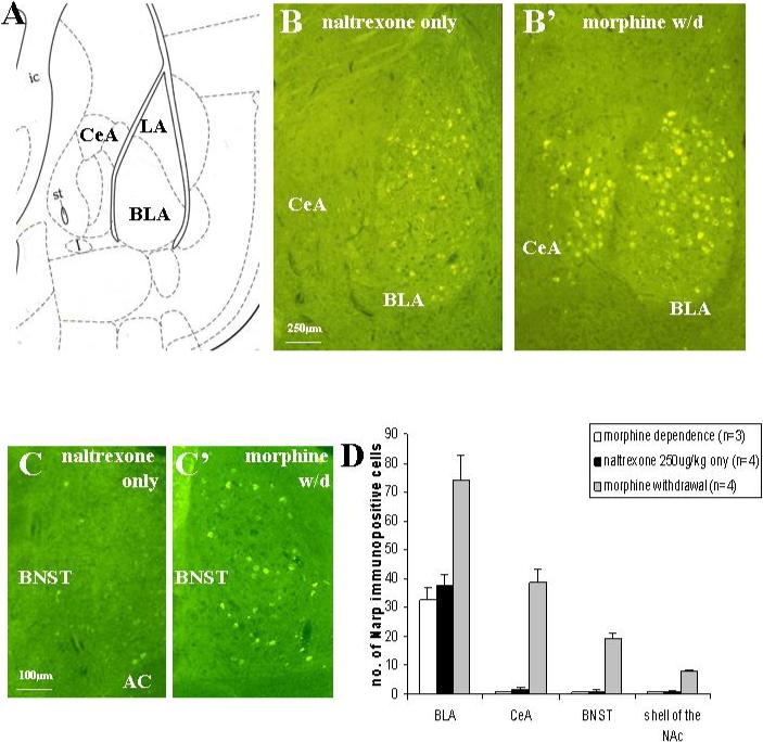
Figure 2.

Narp immunoreactivity in the mouse amygdala is induced by morphine withdrawal. Panel (A) shows an outline of the BLA and CeA from figure 42 of the Paxinos and Franklin (2001) mouse atlas. (B) Constitutive Narp cell body staining is present in the BLA of a morphine-naive mouse treated with naltrexone 250 μg/kg sc, but Narp cell body staining is not detected in either the CeA or lateral amygdala. Four hours following administration of naltrexone 250 μg/kg sc to morphine dependent mice, Narp cell body staining is induced in both the BLA and the lateral portion of the CeA. Panel (C) shows induction of Narp staining in the BNST four hours following morphine withdrawal. (D) Narp cell body staining quantification. ANOVA comparing staining in the BLA and extended amygdala following morphine withdrawal with staining after naltrexone given to morphine-naïve animals and saline given to morphine-dependent animals. BLA: F(2,8)=12.17, p<0.005; after Bonferroni correction, number of Narp-positive cells after morphine withdrawal is significantly greater than both controls (p<0.01). CeA: F(2,8)=56.95, p<0.001; after Bonferroni correction, number of Narp-positive cells after morphine withdrawal is significantly greater than both controls (p<0.001). BNST: F(2,6)=70.22, p<0.001; after Bonferroni correction, number of Narp-positive cells after morphine withdrawal is significantly greater than both controls (p<0.001) NAc: F(2,6)=115.8, p<0.001; after Bonferroni correction, number of Narp-positive cells after morphine withdrawal is significantly greater than both controls (p<0.001). (Note: for BNST and NAc staining only 3 morphine withdrawal and 3 naltrexone only animals were examined.) Narp cell body staining levels in BLA and extended amygdala of morphine-naïve mice treated with naltrexone and morphine-dependent animals given saline is comparable to levels in naïve mice or naïve mice given saline only.
