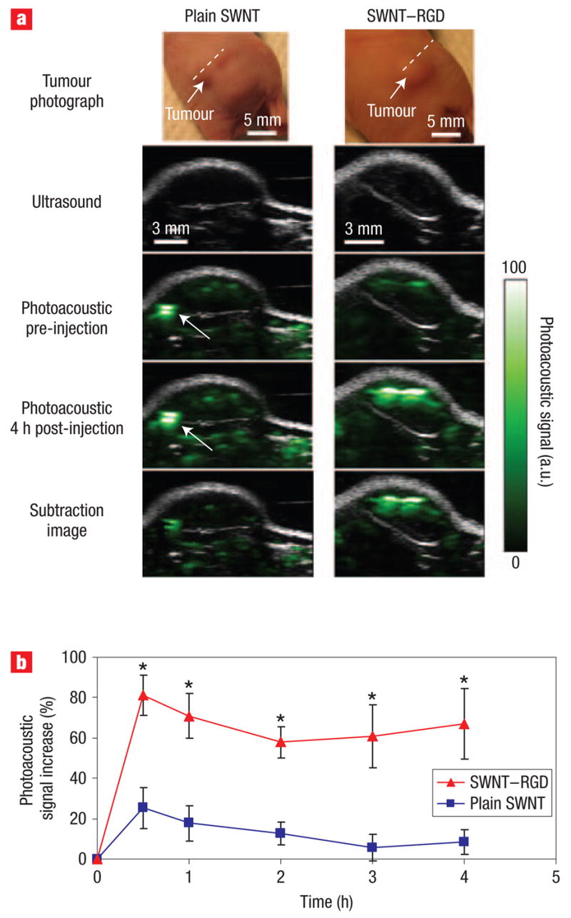
Figure 3. Single-walled carbon nanotube targets tumour in living mice.

a, Ultrasound (grey) and photoacoustic (green) images of one vertical slice (white dotted line) through the tumour. The ultrasound images show the skin and tumour boundaries. Subtraction images were calculated as the 4 h post-injection image minus the pre-injection image. The high photoacoustic signal in the mouse injected with plain single-walled carbon nanotubes (indicated with a white arrow) is not seen in the subtraction image, suggesting that it is due to a large blood vessel and not single-walled carbon nanotubes. b, Mice injected with SWNT –RGD showed a significantly higher photoacoustic signal than mice injected with plain single-walled carbon nanotubes (P < 0.001). The error bars represent standard error (n = 4). *P < 0.05.
