
Figure 5.
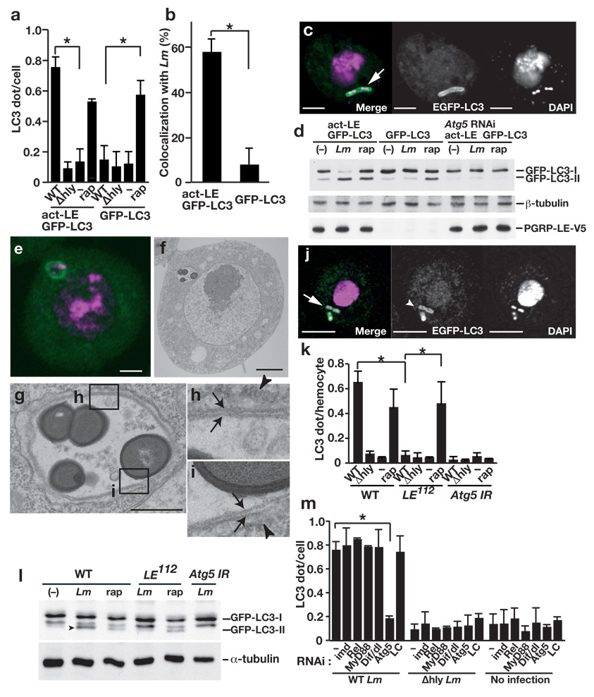
PGRP-LE mediates autophagosome formation in S2 cells and hemocytes. (a) The number of dot- or ring-shaped GFP-LC3 signals per cell was quantified after wild-type (WT) or Δhly L. monocytogenes infection or without infection (−) of S2 cells expressing both PGRP-LE and GFP-LC3 or only GFP-LC3, or after 1.5 h incubation with 5 µM rapamycin (rap). Bars indicate standard deviation of triplicate measurements. (b) The number of GFP-LC3 dots colocalized with wt L. monocytogenes in indicated S2 cells was quantified using confocal microscopy images. Bars indicate standard deviation of triplicate measurements. (c) Confocal microscopy images of WT L. monocytogenes infected S2 cells expressing PGRP-LE and GFP-LC3. GFP-LC3 (green), DAPI (magenta). Scale bar, 5 µM. Arrow indicates co-localization of GFP-LC3 and L. monocytogenes. (d) Indicated S2 cells were infected with WT L. monocytogenes (Lm), or treated with 5 µM rapamycin (rap) and indicated proteins in lysates were detected by immunoblotting. (e–i) Ultrastructural analysis of WT L. monocytogenes-infected S2 cells expressing PGRP-LE and GFP-LC3. (e,f) Fluorescence microscopy (e) and electon microscopy (f) images of cells expressing GFP-LC3 (green) stained with DAPI (magenta). Scale bars, 1 µm. (g) Magnified image of a bacteria-containing vacuole shown in (f). Scale bar, 500 nm. (h, i) Magnified images of the fields indicated in (g). Arrows indicate double-membrane structure that surrounds the bacteria. Arrowheads indicate endoplasmic reticulum–like membrane. (j) Confocal microscopy images of WT L. monocytogenes-infected hemocytes. GFP-LC3 (green), DAPI (magenta). (k) The number of dot- or ring-shaped GFP-LC3 signals per cell in ex vivo-cultured hemocytes expressing GFP-LC3 infected or treated as indicated. Bars indicate standard deviation of triplicate measurements. (l) Hemocytes from third instar larvae of the indicated genotype were cultured ex vivo and infected or treated as indicated and lysates were probed with antibodies specific for the indicated antibodies. Arrowhead indicates processed form of GFP-LC3. (m) S2 cells expressing PGRP-LE and GFP-LC3 under the control of an actin promoter were transfected with double-stranded RNA specific for indicated transcripts (RNAi, below graph) and infected with L. monocytogenes for 0.5 h. After 1 h incubation in gentamicin-containing medium, GFP-LC3 dot formation was quantified by confocal microscopy. Bars indicate standard deviation of at least triplicate measurements. * P < 0.001 (t-test).