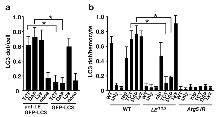
Figure 6.

PGRP-LE is responsible for TCT and DAP-type PGN-induced autophagy. (a) S2 cells expressing PGRP-LE (act-LE) and GFP-LC3, or GFP-LC3 only, were treated with 100 nM TCT, 100 µg/ml highly purified DAP-type peptidoglycans from L. plantarum (DAP), or lysine-type peptidoglycans from S. epidermidis (Lys). After 2 h incubation, GFP-LC3 dot formation was quantified. Bars indicate the variance of two independent experiments. (b) The number of dot- or ring-shaped GFP-LC3 signals per hemocyte of indicated genotype was quantified after wild-type (WT) or Δhly L. monocytogenes infection, 5 µM rapamycin treatment (rap), or treatment with 100 nM TCT (TCT), 100 µg/ml highly purified DAP-type PGN from L. plantarum (DAP), or lysine-type PGN from S. epidermidis (Lys). Bars indicate variance of two independent experiments. Genotypes: w−; UAS-GFP-LC3/UAS-Gal4; heat-shock-GAL4/+ (WT), PGRP-LE112 ; UAS-GFP-LC3/UAS-Gal4; heat-shock-GAL4/+ (LE112), UAS-Atg5 IR/+, UAS-GFP-LC3/UAS-Gal4; heat-shock-GAL4/+ (Atg5 IR). * P < 0.001 (t-test).
