
Fig. 6.
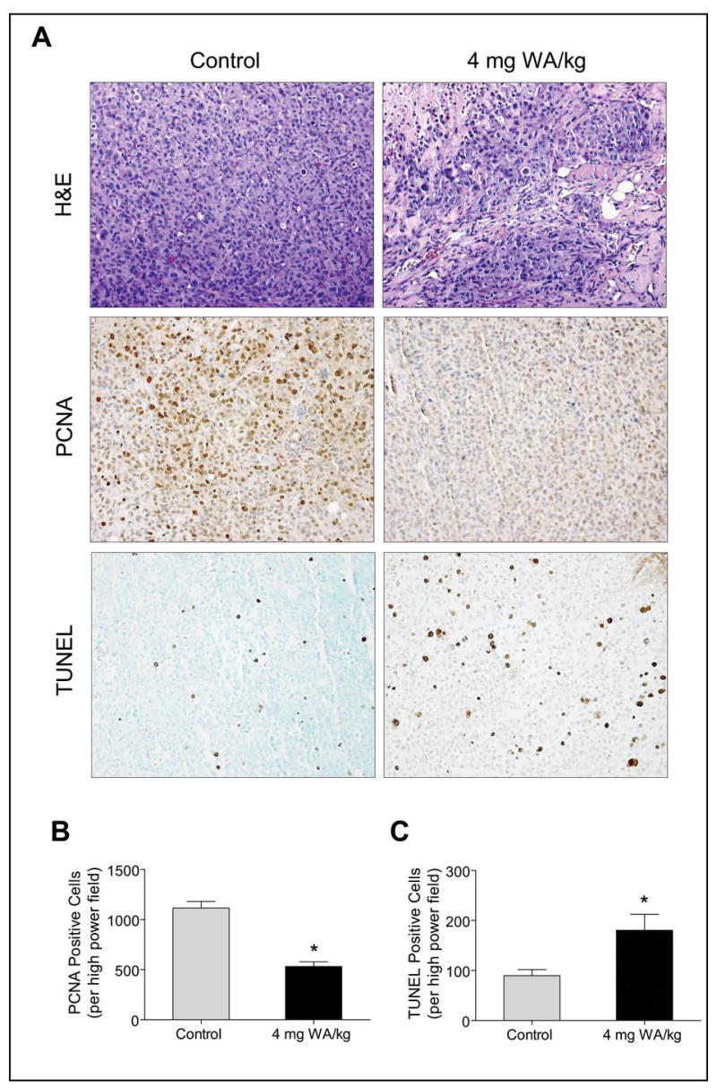
A, histological analysis of MDA-MB-231 tumors from control and WA-treated mice (subcutaneous xenograft study) for H&E staining, proliferating cell nuclear antigen (PCNA) expression, and TUNEL positive apoptotic bodies. Representative H&E, PCNA, and TUNEL staining in tumor sections of a control and a WA-treated mouse are shown. B, quantitation of PCNA expression in tumors from control and WA-treated mice. C, quantitation of apoptotic bodies/high-power field in tumors from control and WA-treated mice. At least three randomly selected fields on each slide from tumors of five individual mouse of control and WA-treated groups were scored for PCNA expression and TUNEL positive cells. Columns, mean (n= 5), bars, SE. *P<0.05, significantly different compared with control by t-test.