
Figure 5.
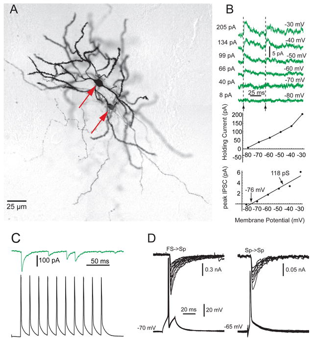
Feedback inhibition between spiny neurons. A. Synaptically connected pair of spiny neurons (arrows) in a striatal slice from an adult rat stained with biocytin after whole cell recording. B. Sp-Sp IPSCs recorded in perforated patch configuration in a slice from a 13 day old rat. Top. Presynaptic spikes were triggered at the arrows while holding the postsynaptic cell at the membrane potentials indicated on the right of the traces. Middle. Plot of the current required to hold the membrane at different potentials between -80 and -30 mV. Bottom. Slope of the VI plot from the data at top shows the conductance change due to the collateral synapse is only 118 pS. The amplitude goes to zero at the reversal potential of -76 mV. C. Response to a train of presynaptic spikes at 40 Hz shows frequent failures. D. Comparison of whole cell recordings of IPSCs in two different spiny cells, one elicited by single spikes in an FS interneuron (left) and another in a spiny cell (right). Note the difference in the voltage calibration and the approximately 6-fold difference in maximum IPSC amplitude. The internal contained 140 mM CsCl. Redrawn from Koós et al., (2004). Copyright 2004 by the Society for Neuroscience.