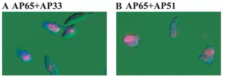
Fig. 1.
Demonstrations of surface colocalization of AP65 with either AP33 (A) or AP51 (B) by fluorescence using non-permeabilized trichomonads of isolate T016 grown in a high-iron medium as described in Experimental procedures. Organisms were treated with antiserum IgG to adhesins conjugated individually to fluorescein (anti-AP65) or rhodamine (both anti-AP51 and AP33). Use of the individual antisera gave only the respective colour, and blending was observed indicative of coexpression. Little or no fluorescence was observed with prebleed NRS or with trichomonads grown in a low-iron medium (Lehker et al., 1991; Arroyo et al., 1992). In this experiment, fluorescence was examined with an Axiophot II Zeiss epifluorescent microscope.

