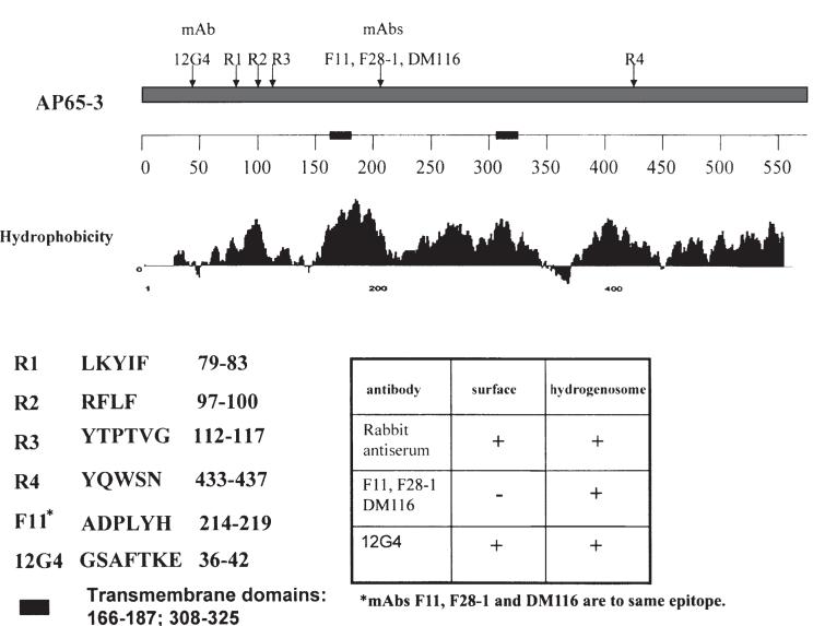
Fig. 3.
Identification of epitopes using overlapping decapeptides obtained for AP65-3 as described in the Experimental procedures section. The IgG1 mAb F11 and two new IgG1 mAbs, F28-1, and DM116 generated from distinct mAb libraries recognized the same epitope. A LamB-AP65 fusion protein used for immunization of mice resulted in generation of mAb 12G4 toward an epitope at the amino terminus of the clustered rabbit epitopes. The sequences of the epitopes are included on the left of the insert table, which presents the combined fluorescence and immunogold labelling results of separate experiments. The new mAb reacted with the parasite surface as shown in Fig. 4. Two transmembrane domains are represented on the numbered scale by bold, solid bars, and the amino acid numbers are indicated.

