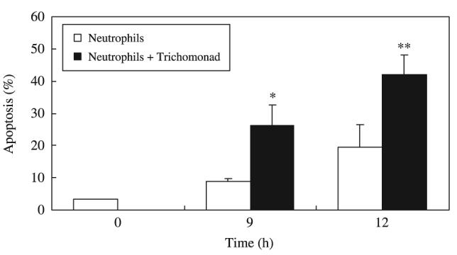
Figure 3.

Time course of apoptosis of human neutrophils co-incubated with Trichomonas vaginalis. Neutrophils (1 × 10⁶/mL) were co-cultured with live T. vaginalis (1 × 10⁵/mL) for 0-12 h. The percentage of apoptotic cells was obtained by flow cytometry using DiOC6. The data represent mean ± SEM of four separate experiments. *P < 0·05; **P < 0·01, Neutrophils vs. Neutrophils + T. vaginalis.
