
Figure 3. Drosophila model of tauopathy.
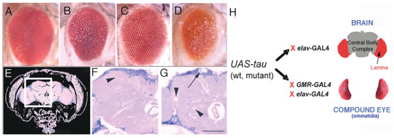
(A–D) The tau-induced rough eye phenotype consists of a small irregular eye with fused ommatidia and small focal areas of degeneration. Compare the elav-GAL4/+ (A) and GMR-GAL4/+ (C) control eyes with eyes of transgenic flies expressing mutant forms of tau, tauR406W (B; genotype: elav-GAL4/+; UAS-tauR406W6/+) or tauV337M (D; genotype: UAS-tauV337M1/+; GMR-GAL4/+), This phenotype is visible in adults at eclosion. (E) Hematoxylin and eosin-stained frontal section through the head and brain of a wild-type fly. (F–G) Areas of the fly brain similar to that boxed in (G) are shown for control elav-GAL4/+ (F) and mutant tau-expressing (G) flies. Vacuolization is observed in the neuropil (N, arrowheads) and cortex (Cx, arrow) [42]. (H) Bipartite UAS/GAL4 system: This system allows for specific expression of tau either in postmitotic neurons, including the fly brain, with elav-GAL4, or in the Drosophila eye with elav-GAL4 (photoreceptors only) or GMR-GAL4 (photoreceptors and support cells). F and G have been reproduced with permission from [42].