
Figure 2. Correlation of Jagged-1 immunoreactivity with nuclear localization of Notch3 in ovarian carcinomas.
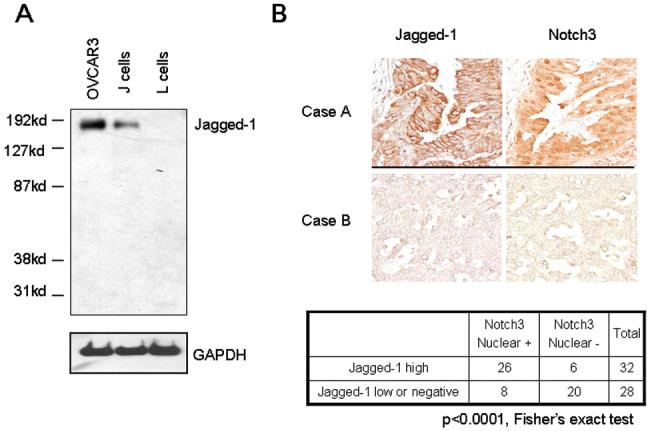
A. Jagged-1 protein expression was determined by Western blot in OVCAR3, J, and L cells to validate the antibody specificity. J cell is an Ltk- mouse fibroblast cell line that was engineered to express the full-length Jagged-1. L cell is the parental Ltk- cell line. Jagged-1 protein bands (∼175 kD) were detected in both OVCAR3 and J cells but not in L cells. B. Upper panel: Immunoreactivity of Jagged-1 and Notch3 in two representative ovarian high-grade carcinoma tissues. Case A shows intense Jagged-1 membrane staining and prominent Notch3 nuclear immunoreactivity. In contrast, case B shows very weak Jagged-1 and Notch3 staining. Lower panel: Summary table of Jagged-1 and Notch3 immunohistochemistry for 60 high-grade carcinomas. Jagged-1 protein expression and nuclear localization of Notch3 is significantly correlated (p<0.0001, Fisher’s exact test).