
Figure 4. Growth analysis of parental SLE and chimeric viruses in Vero cells or in human neuroblastoma cells following incubation at different temperatures.
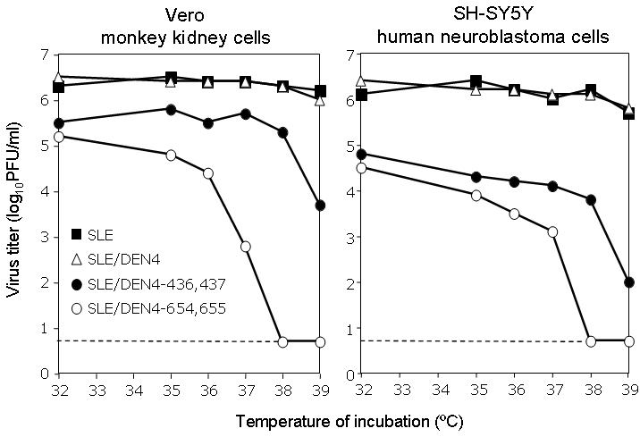
An efficiency of plaque formation assay was performed with the indicated viruses in Vero cells and SH-SY5Y cells. Confluent monolayers of cells were infected with serial ten-fold dilutions of virus at 32°C, overlaid with semisolid growth media, and then incubated for five days at 32, 35, 36, 37, 38, or 39°C. Plaques were visualized by immunostaining and quantitated. The limit of detection (100.7 PFU/ml) is indicated by a dashed line.