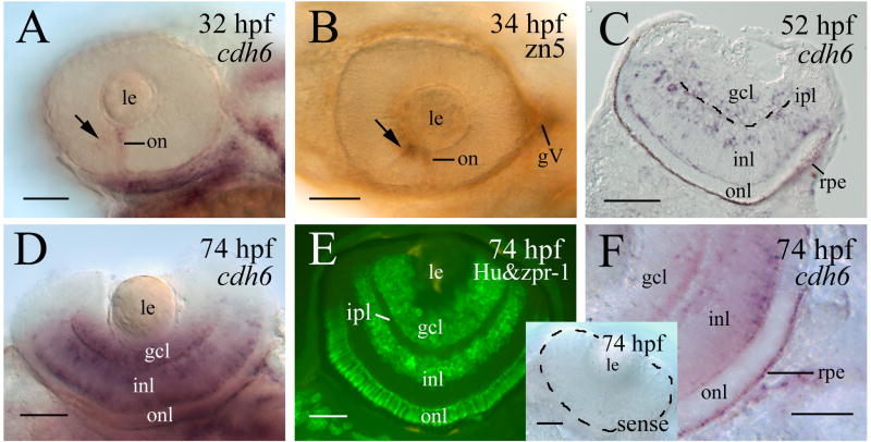
Figure 1.
cdh6 expression in the developing zebrafish retina. Panels A, B and D show whole mount eyes (anterior to the left, dorsal is up for panels A and B, and dorsal is down for panel D). Panels A, D and F are whole mount eyes processed for cdh6 in situ hybridization. Panel B is a whole mount eye processed for zn5 immunostaining (labeling early differentiating retinal ganglion cells). Panel C is a retinal cross section (dorsal to the left) of an embryo processed for cdh6 whole mount in situ hybridization, while panel E is immunostaining of a retinal cross section (dorsal to the left) using anti-HuC/HuD (labeling the retinal ganglion cells and amacrine cells) and zpr-1 antibodies (labeling the photoreceptor layer). Arrows in panels A and B indicate labeling in the anteroventral region of the retina. The inner plexiform layer (ipl) in panel C is indicated by the dashed line. Panel F is a higher magnification of the posteroventral quadrant of a whole mount eye (anterior to the left and dorsal up) showing labeling in the retinal pigmented epithelium (rpe). The insert in panel F shows a whole mount eye (outlined by the dashed line) processed for in situ hybridization using a cdh6 sense probe. Other abbreviations: gcl, retinal ganglion cell layer; gV, trigeminal ganglion; inl, inner nuclear layer; le, lens; on, optic nerve; onl, outer nuclear layer. Scale bars = 50 μm.

