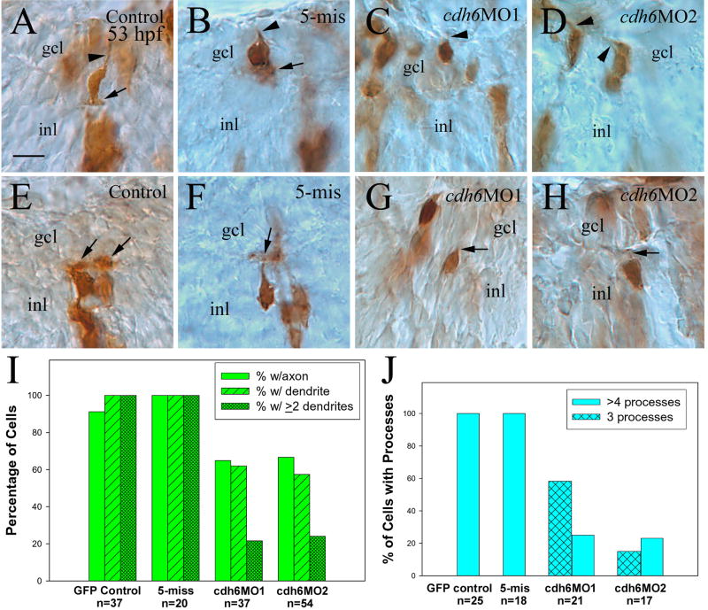
Figure 10.
Analysis of cadherin-6 function in differentiation of individual RGCs (panels A-D, and I) and amacrine cells (panels E-H, and J) labeled with an anti-eGFP antibody. All images are from cross sections (30 μm). Arrowheads point to axons, while arrows indicate dendrites. The number (n) in panels I and J represents the number of retinal cells examined for each group. Abbreviations are the same as in Figure 1. Scale bar = 10 μm.

