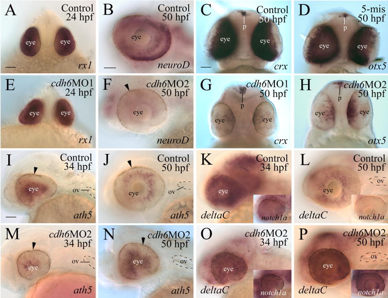
Figure 6.
Expression of transcription factors and Notch-Delta genes in the control and cdh6 morphant retinae. Panels A, C, D, E, G and H are in-face views (dorsal up) of embryo heads from embryos processed for whole mount in situ hybridization. The remaining panels are lateral views (anterior to the left and dorsal up) of whole mount eyes and/or heads. The arrowhead in panels F, I, J, M and N points to retinal pigmented epithelium. Abbreviations: p, pineal gland; ov, otic vesicle. Panels A and E, B and F, C, D, G and H are of the same magnifications, respectively. Panels I-P are of the same magnifications. Scale bars = 50 μm.

